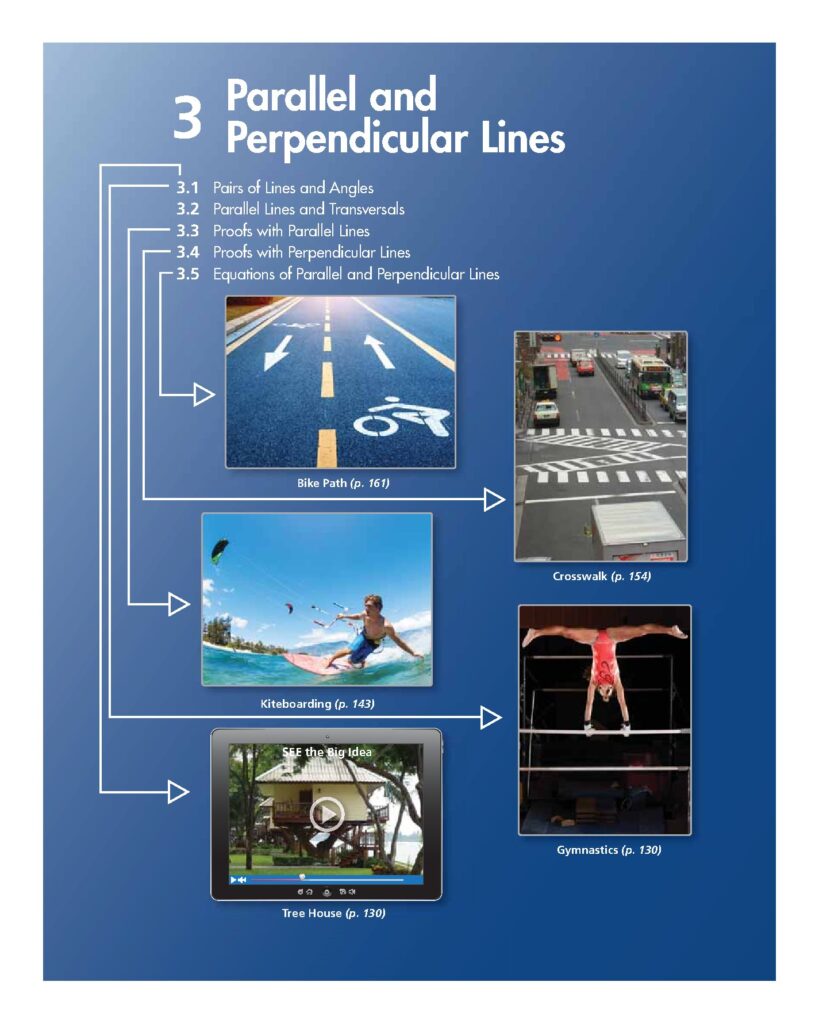
Bones of the Gluteal Region Practical Session 2017
The 'Bones of the Gluteal Region' practical session from 2017 provides an in-depth exploration of the hip bone's anatomy, including the ilium, ischium, and pubis. It emphasizes the anatomical position of the pelvis and its significance in understanding how structures transition from the pelvis to the thigh. The session includes detailed diagrams and X-ray images to illustrate the development and articulation of the hip bones. This resource is essential for students studying human anatomy, particularly those focusing on the pelvic girdle and its related structures.
Key Points
Explains the anatomical position of the hip bone and its importance.
Includes diagrams illustrating the hip bone's structure and articulation.
Covers the development of the triradiate cartilage and its ossification process.
Details the relationship between the pelvis and thigh structures, including arteries and nerves.
This link leads to an external site. We do not know or endorse its content, and are not responsible for its safety. Click the link to proceed only if you trust this site.
FAQs of Bones of the Gluteal Region Practical Session 2017
What are the main components of the hip bone?
The hip bone consists of three main components: the ilium, ischium, and pubis. The ilium is the superior part, while the ischium is located posteriorly and inferiorly, and the pubis is positioned anteriorly and inferiorly. Understanding these components is crucial for studying the pelvic girdle and its functions.
How does the anatomical position of the pelvis affect movement?
The anatomical position of the pelvis is critical as it determines how structures from the pelvis connect to the thigh. When the pelvis is oriented forward, it facilitates the proper alignment of muscles, nerves, and blood vessels that transition from the pelvic region to the thigh, impacting mobility and stability.
What is the significance of the triradiate cartilage?
The triradiate cartilage is significant because it is the area where the ilium, ischium, and pubis meet at the acetabulum. This cartilage plays a crucial role in the development of the hip bone, as it ossifies over time, particularly around the age of 17, leading to a bony union that is essential for structural integrity.
What role do the sacroiliac joints play in the pelvis?
The sacroiliac joints connect the hip bones to the sacrum, providing stability and support to the pelvis. These joints allow for limited movement, which is essential for absorbing shock during activities such as walking and running. Understanding their function is vital for studying lower limb biomechanics.
What muscles attach to the hip bone?
Several important muscles attach to the hip bone, including the gluteus maximus, gluteus medius, and tensor fasciae latae. These muscles are crucial for hip movement, stability, and overall lower body function. Their attachment points on the hip bone are essential for understanding muscle mechanics and anatomy.
Related of Bones of the Gluteal Region Practical Session 2017