Understanding The Eukaryotic Cell Cycle A Biological And Experimental Overview

Understanding The Eukaryotic Cell Cycle A Biological And Experimental Overview

The eukaryotic cell cycle is a complex process essential for cellular replication and division. This overview explores the phases of the cell cycle, including G1, S, G2, and M phases, and highlights the critical checkpoints that ensure genomic integrity. Key molecular mechanisms, such as the roles of cyclins and cyclin-dependent kinases (Cdks), are discussed in detail. This resource is invaluable for biology students and researchers interested in cell biology and cancer studies, providing insights into DNA replication and cell proliferation biomarkers. It also outlines various experimental techniques for assessing cell cycle kinetics.

Key Points

  • Explains the phases of the eukaryotic cell cycle, including G1, S, G2, and M phases.
  • Discusses the roles of cyclins and cyclin-dependent kinases (Cdks) in cell cycle regulation.
  • Highlights key biomarkers for cell proliferation, such as Ki-67 and PCNA.
  • Details molecular techniques for assessing DNA replication and cell cycle kinetics.
311
/ 8
Bio-Rad
Laboratories, Inc.abdserotec.com
LIT.NPA.2013.1 © Copyright Bio-Rad Laboratories, Inc. All rights reserved. Published by AbD Serotec, a Bio-Rad Company, Endeavour House, Langford Lane, Langford Business Park,
Kidlington, OX5 1GE. AbD Serotec reagents are for research purposes only, not for therapeutic or diagnostic use. Cy containing products or portions thereof are manufactured under license
from Carnegie Mellon University under U.S. Patent Number 5,268,486 and related patents. C
y
®
and CyDye
®
are registered trademarks of GE Healthcare Limited. Alexa Fluor
®
is a registered
Contents
1. Overview of the Cell Cycle in Eukaryotes
1.1 Introduction to the Eukaryotic Cell Cycle
1.2 Cell Cycle Control
1.3 DNA Replication in Eukaryotes
1.4 Key Cell Proliferation Biomarkers
2. Molecular Techniques for Assessing DNA Replication and Cell Cycle Kinetics
2.1 Assessing DNA Replication
2.2 Quantifying DNA Content
2.3 Measuring the Expression of Cyclins and Cdks
2.4 Detecting Cell Proliferation Markers
2.5 Summary and Future Outlook
1. Overview of the Cell Cycle in Eukaryotes
From as early as the 19th century, scientists have been intensely investigating the cell cycle. During this period, they
discovered that new cells are derived from pre-existing cells (Nurse et al. 1998). However, the exact processes involved
in cell division were largely unknown. Of particular scientific interest was to determine how the process differed among
species.
In the last 60 years, there have been significant discoveries and insights into the molecular mechanisms involved in cell
division in prokaryotes and eukaryotes. These studies demonstrated that the cell cycle in eukaryotes is much more
complex than in prokaryotic organisms. Although, DNA replication and cell division occur in both cases, the processes
vary significantly. A main difference lies in how these organisms replicate their DNA. Since the average eukaryotic
cell has 25 times more DNA than prokaryotic cells, prokaryotic cell division does not include DNA condensation into
chromosomes as observed in the eukaryotic cell cycle. Another major difference lies in the stages at which DNA
replication occurs. Prokaryotes replicate their DNA continuously throughout their relatively short cell cycle whereas
eukaryotic cells replicate DNA only in the S-phase of the cell cycle (discussed in detail in Section 1.2).
This mini-review will specifically discuss the intricacies of the eukaryotic cell cycle with special focus on DNA replication,
cell cycle control, and key biomarkers of cell division.
The eukaryotic cell cycle is an evolutionarily conserved process that results in the replication of cells. It is tightly
regulated, and includes three major checkpoints: G1, G2/M, and spindle (M). These checkpoints monitor the
order, fidelity, and integrity of each phase of the cell cycle. For example, the G2/M checkpoint detects potential
DNA damage, thus allowing repair before cell division. Defects in cell cycle progression often result in diseases
such as cancer. Accordingly, this essential life cycle is routinely used to assess cell health, as well as for cancer
prognosis and diagnosis. This mini-review provides an overview of the eukaryotic cell cycle, including common
molecular techniques for evaluating proliferating cells.
Cancer
Understanding the Eukaryotic Cell Cycle — a Biological and
Experimental Overview
Mini
Review
2
1.1 Introduction to the Eukaryotic Cell Cycle
The highly regulated cell cycle is divided into phases,
referred to as interphase (G1, S, and G2) and the mitotic
(M) phase (Figure 1). In the gap 1 (G1) phase, the cell grows
and acquires the energy needed for division. Cellular
components, except for chromosomes, are duplicated
at this stage. In the synthesis (S) phase, DNA replication
occurs to duplicate the genetic material, with each
chromosome now consisting of two sister chromatids.
In the gap 2 (G2) phase, the cell prepares to divide by
inducing metabolic changes that assemble the cytoplasmic
components necessary for mitosis. During the M phase,
nuclear division occurs, and the cell finally divides to
create two identical daughter cells. The physical process
of creating two daughter cells through division of the
nucleus, cytoplasm, and plasma membrane is referred to as
cytokinesis (derived from the Greek kyto or kýtos meaning
container, body, or receptacle and kinesis meaning
movement). At the end of cytokinesis each new cell consists
of a full complement of DNA from the parent cell (Kapinas
et al. 2013).
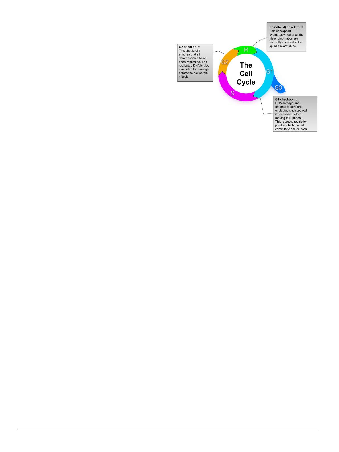
Fig. 1. Overview of the eukaryotic cell cycle. During cell division, cells
pass through a series of stages collectively referred to as the cell cycle. To
ensure that healthy cells are produced after each round of cell division, the cell
cycle consists of three major checkpoints with distinct functions: G1, G2, and
Spindle (M) checkpoints.
Another specialized cell division process known as meiosis is required to produce egg and sperm cells for reproduction.
This process is split into meiosis I and meiosis II, in which meiosis I is unique to germ cells and meiosis II is similar to
mitosis. However, in contrast to mitosis, the molecular and regulatory mechanisms involved in meiosis are less understood
(Ohkura 2015).
Under certain conditions, a cell can exit the cell cycle and enter a state of quiescence referred to as the gap 0 (G0) phase.
This phase is however reversible and G0 cells can return to the G1 phase and resume growth and division if appropriately
stimulated.
1.2 Cell Cycle Control
Each phase of the cell cycle is tightly regulated, with checkpoints in place near the end of G1, at the G2/M transition, and
near the end of the metaphase stage of mitosis (spindle (M) checkpoint). These checkpoints are surveillance mechanisms
whose function is to ensure that the generated daughter cells are duplicates of the parent cell complete with the accurate
number of chromosomes and are mutation free (Figure 1). During the G1 checkpoint, cellular conditions necessary for
progression through the cell cycle are evaluated. A cell generally passes the G1 checkpoint if it is an appropriate size,
possesses adequate energy, and does not have damaged DNA. The main function of the G2 checkpoint is to ensure
that replication of all chromosomes is complete and without introductions of mutations or unrepaired DNA damage. In
addition, appropriate cell size and protein reserves are also assessed during this checkpoint. The spindle/M checkpoint
ensures that all sister chromatids are correctly attached to the spindle microtubules and that each cell has the correct
number of chromosomes.
These checkpoints halt cell cycle progression if the cell has not met each of the requirements being evaluated. This is
necessary to allow the identified unfavorable conditions to be addressed. For example, detected DNA damage leads to
the activation of the p53 transcription factor, which has been referred to as the ‘guardian of the genome’ due to its major
role in maintaining genome stability (Lane 1992). The main function of p53 is to induce cell cycle arrest at the G1 or G2/M
phases and initiate DNA repair. It activates gene expression of DNA repair genes such as P53R2 (Tanaka et al. 2000). p53
can also induce apoptosis as a last resort, if the damaged DNA cannot be repaired, by inducing expression of apoptotic
genes such as BAX (Zilfou and Lowe 2009). Since it plays such an important role in preventing the continued cell cycle
progression of cells with mutated DNA, p53 is considered a tumor suppressor (Zilfou and Lowe 2009). Consequently, it
has been reported to be commonly mutated or absent in several types of cancer (Hussain and Harris 1998).
The master regulators of the cell cycle in eukaryotes are however heterodimeric enzyme complexes, which consist of
cyclins and cyclin-dependent kinases (Cdks) (Murray 2004). The expression of cyclins increases or decreases in distinct
phases of the cell cycle, and they are divided into groups based on the cell cycle phase that they regulate (Figure 2)
(Murray 2004). However, in most cases, the concentration of Cdks remains relatively constant. Each Cdk subunit can
associate with different cyclins, and the associated cyclin determines which protein substrates are phosphorylated by the
Cdk-cyclin complex (Lodish et al. 2000). Moreover, Cdks have no kinase activity unless cyclin bound. In addition to the
3
binding of cyclins, activation of the complex also requires phosphorylation of key residues in the activation loop of the Cdk
subunit (Harper and Elledge 1998, Hochegger et al. 2008).
Several mechanisms have been identified for inhibiting activated cyclin-Cdk complexes. These include inhibitory
phosphorylation of important residues such as tyrosine 15 and threonine 14 in Cdk1, degradation of the cyclin subunits by
specific ubiquitin-mediated proteolysis, or association of the complex with a highly specific inhibitor protein such as p16 in
the case of the cyclin D-Cdk4 complex (Hochegger et al. 2008, Kellogg 2003, Peters 2006, Serrano et al. 1993).
Fig. 2. Expression of cyclins throughout the cell cycle phases (Lodish et al. 2000). Cyclins are differentially expressed at various phases of the cell cycle and play
distinct roles in cell cycle control. The figure demonstrates the stages in the cell cycle in which each cyclin is expressed. The grey shaded areas represent the peak
expression of the respective cyclin.
The classical model of cell cycle control indicates that D-type cyclins and Cdk4 or Cdk6 regulate events in the early
G1 phase (Nurse 2000). The cyclin E-Cdk2 complex then initiates the S phase and cyclin A-Cdk2 or cyclin A-Cdk1
complexes regulate the completion of the S phase. The cyclin B-Cdk1 complex is subsequently responsible for mitosis
(Table 1). The transition between each phase of the cell cycle is mediated by protein phosphorylation, which is catalyzed
by Cdks. In response, the removal of phosphate residues by phosphatases is also critical for cell cycle progression. For
example, several phosphatases are involved in the control of mitosis (Chen et al. 2007). Protein phosphatase-2A1 (PP2A1)
mediates the main phosphatase activity towards mitotic substrates (Sola et al. 1991). PP2A is deactivated when cells enter
mitosis but is reactivated after the proteolysis of mitotic cyclins (Sola et al. 1991).
A decade ago, a revised model of eukaryotic cell cycle regulation, called the minimal threshold model, was proposed
(Hochegger et al. 2008). This model stipulates that either Cdk1 or Cdk2 bound to cyclin A is sufficient to control all stages
of interphase, whereas the cyclin B-Cdk-1 complex is necessary for the transition to mitosis (Hochegger et al. 2008).
It also postulates that the differences between interphase and mitotic Cdks are not necessarily related to the specific
cyclins they interact with. However, this is due to localization and a higher activity threshold for mitosis than interphase
(Hochegger et al. 2008).
Knock out mice provide data in support of this model demonstrating that deletion of certain Cdks and cyclins does
not lead to disruption of the cell cycle in somatic cells (Berthet and Kaldis 2007, Hochegger et al. 2008, Malumbres
and Barbacid 2005). For example, the embryos of mice lacking Cdk2, Cdk4, and Cdk6 still carry out a functional cell
cycle (Santamaria et al. 2007). However, according to the classical view, cells from these mice should not be able to
progress beyond the G1 phase (Hochegger et al. 2008). This suggests that only a few complexes such as Cdk1-cyclin A
are necessary for cell cycle progression. The minimal threshold model is also supported by other studies in yeast and
mathematical modeling experiments (Coudreuse and Nurse 2010, Gérard et al. 2015). Further studies are however
needed to confirm this proposed model, such as those that provide an explanation for the differential effects of Cdk
deletions. According to Hochegger et al. (2008), this could be due to yet unknown kinase-independent functions of
Cdk-cyclin complexes.
/ 8
End of Document
311
You May Also Like

FAQs of Understanding The Eukaryotic Cell Cycle A Biological And Experimental Overview

What are the main phases of the eukaryotic cell cycle?
The eukaryotic cell cycle consists of several key phases: G1 (gap 1), S (synthesis), G2 (gap 2), and M (mitosis). In the G1 phase, the cell grows and prepares for DNA replication. The S phase is where DNA replication occurs, resulting in two sister chromatids for each chromosome. During the G2 phase, the cell prepares for division, ensuring all cellular components are ready. Finally, in the M phase, the cell undergoes mitosis, resulting in two identical daughter cells.
How do cyclins and Cdks regulate the cell cycle?
Cyclins and cyclin-dependent kinases (Cdks) are crucial for regulating the cell cycle. Cyclins are proteins whose levels fluctuate throughout the cell cycle, activating Cdks when bound. This activation leads to the phosphorylation of target proteins, driving the cell through various checkpoints. For instance, cyclin D and Cdk4 regulate the transition from G0 to G1, while cyclin B and Cdk1 are essential for the onset of mitosis. This tightly controlled mechanism ensures proper cell division and genomic stability.
What are some key biomarkers of cell proliferation?
Key biomarkers of cell proliferation include Ki-67, PCNA, and MCM2. Ki-67 is a nuclear protein that is present during all active phases of the cell cycle but absent in resting cells. PCNA plays a vital role in DNA replication and repair, while MCM2 is essential for the initiation of DNA replication. The expression levels of these biomarkers can indicate cell growth and are often used in cancer diagnostics to assess tumor proliferation rates.
What techniques are used to assess DNA replication in the cell cycle?
Several techniques are employed to assess DNA replication during the cell cycle. One common method involves using thymidine analogs, such as BrdU, which are incorporated into newly synthesized DNA. These analogs can be detected using specific antibodies, allowing researchers to quantify DNA synthesis. Other methods include flow cytometry with fluorescent dyes that bind to DNA, enabling the measurement of DNA content across different cell cycle phases. These techniques are crucial for understanding cell proliferation and the effects of various treatments.

Related of Understanding The Eukaryotic Cell Cycle A Biological And Experimental Overview