Gizmos Student Exploration: Cell Types

Gizmos Student Exploration: Cell Types

Cell Types exploration focuses on understanding various cell types through microscopy. Students will observe human skin, plant cells like Elodea, and unicellular protists, enhancing their knowledge of cellular structures and functions. This resource is ideal for biology students aiming to grasp cellular biology concepts and prepare for exams. The exploration includes detailed observations, comparisons of animal and plant cells, and insights into specialized cell functions. Engaging with this material will help students develop critical thinking skills in biological sciences.

Key Points

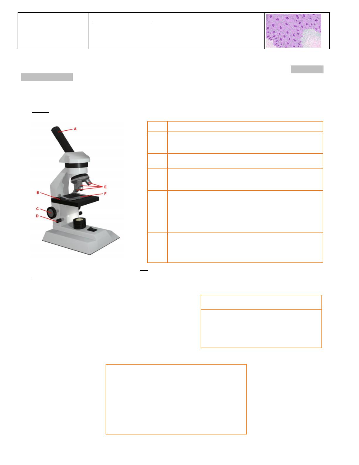
  • Explores human skin cells and their structures using a microscope.
  • Covers plant cell observations, including Elodea and maple leaf cells.
  • Includes detailed comparisons of animal and plant cells for better understanding.
  • Examines specialized cells like neurons and their functions in the human body.
145
/ 4
Name:
Date:
Student Exploration: Cell Types
Directions: Follow the instructions to go through the simulation. Respond to the questions and
prompts in the orange boxes.
Vocabulary: ATP, bacteria, carbon dioxide (CO
2
), cell, cellular respiration, compound light microscope, muscle
cell, neuron, organelle, photosynthesis, protist, red blood cell, root hair cell, tissue, white blood cell
Gizmo Warm-up
In the Cell Types Gizmo, you will use a light microscope to compare and contrast different samples. On the
LANDSCAPE tab, click on the Frog Blood. (Turn on Show all samples if you can’t find it.) Switch to the
MICROSCOPE tab to observe the sample as it would appear under the microscope. By default, this
microscope is using 40x magnification.
1. Drag the Coarse focus slider until the sample is focused as well as possible. Then, improve the focus with
the Fine focus slider. What do you see?
2. Select the 400x magnification. Click Show labels. Adjust the fine focus and draw what you see below.
3. What happens to the stage as you drag the Course Focus to the left and right?
___________________________________________________________________________________
4. Which way does the image move when you move the Stage left? _______________________________
5. Which way does the image move when you move the Stage right? _______________________________
6. Which way does the image move when you move the Stage up? _______________________________
7. Which way does the image move when you move the Stage down? _______________________________
Reproduction for educational use only. Public sharing or posting prohibited. © 2020 ExploreLearning™ All rights reserved
Activity A:
Animal cells
Get the Gizmo ready:
On the LANDSCAPE tab, click on the woman’s right
arm to choose the Human skin sample.
Select the MICROSCOPE tab.
Introduction: Complex organisms are made up of smaller units, called cells. Most cells are too small to be
seen by the naked eye. Microscopes are used to magnify small objects, so here you will use a compound
light microscope to observe the cells of different organisms.
Question: What are similarities and differences between cells from different organisms?
1. Match: Read about each microscope part. Match the description to the part on the diagram.
Stage: Platform where a slide is placed.
Eye piece: Lens at the top of the microscope that the
user looks through. This lens most commonly magnifies
a sample by 10x.
Coarse focus knob: Large knob that moves the stage
up and down to focus the sample.
Fine focus knob: Small knob that moves the stage over
a short distance to refine the focus.
Objective lens: A second lens that further magnifies the
sample. Microscopes usually have several objective
lenses with different magnifications. The total
magnification is the product of the eyepiece
magnification and the objective lens magnification.
Slide: A rectangular piece of glass upon which a sample
is mounted for viewing under a microscope.
2. Manipulate: With 40x selected, use the Coarse and Fine focus sliders to focus on the sample. Then,
choose 400x and focus on the sample using the Fine focus slider.
A. Which focus knob is easier to use at 40x? 400x?
B. Turn on Show labels. What structures can you
see in human skin cells?
3. Select the 400x magnification. If necessary, adjust the fine focus. In the space below, draw what you see.
Reproduction for educational use only. Public sharing or posting prohibited. © 2020 ExploreLearning™ All rights reserved
Activity B:
Plant cells
Get the Gizmo ready:
On the LANDSCAPE tab, click on the Elodea leaf sample.
Select the MICROSCOPE tab.
1. Manipulate: With 40x selected, use the Coarse and Fine focus sliders to focus on the sample. Then,
choose 100x and then the 400x and focus on the sample using the Fine focus slider.
2. Turn on Show labels and in the space below, draw what you see..
3. Observe: An organelle is a cell structure that performs a specific function. Observe the samples below
under the highest magnification. Click the Show labels checkbox to label the organelles.
List the organelles in each sample.
Sample
Organelles
Mouse skin
Fly muscle
Maple leaf
Elodea
What do all of these samples have in common?
What organelles are found in the maple leaf and Elodea and not in the mouse or fly?
Reproduction for educational use only. Public sharing or posting prohibited. © 2020 ExploreLearning™ All rights reserved
/ 4
End of Document
145
You May Also Like

FAQs of Gizmos Student Exploration: Cell Types

What types of cells are explored in this Gizmo?
The Gizmo explores various cell types, including human skin cells, plant cells such as Elodea and maple leaf cells, and specialized cells like neurons. Each cell type is observed under a microscope, allowing students to identify key structures and organelles. This exploration helps students understand the similarities and differences between animal and plant cells, as well as the unique functions of specialized cells in multicellular organisms.
How does the Gizmo help students understand cell structures?
The Gizmo provides a hands-on experience where students can manipulate a microscope to observe different cell types. By adjusting magnification and focusing techniques, learners can see the intricate details of each cell, such as the nucleus, cell membrane, and cytoplasm. This interactive approach enhances their understanding of cellular biology and the specific functions of various organelles, making the learning process more engaging and effective.
What are the main differences between plant and animal cells observed?
In the Gizmo, students can observe key differences between plant and animal cells, such as the presence of a cell wall and chloroplasts in plant cells, which are absent in animal cells. Plant cells typically have a more rigid structure due to the cell wall, while animal cells are more flexible. Additionally, the presence of chloroplasts allows plant cells to perform photosynthesis, a function not found in animal cells. Understanding these differences is crucial for grasping the fundamentals of cellular biology.
What role do specialized cells play in the human body?
Specialized cells, such as neurons and muscle cells, perform specific functions that are essential for the body's overall operation. Neurons transmit signals throughout the nervous system, enabling communication between different body parts. Muscle cells are responsible for movement and contraction, allowing for physical activity. The Gizmo highlights how the structure of these cells is adapted to their functions, providing students with a deeper understanding of human biology and the importance of cellular specialization.

Related of Gizmos Student Exploration: Cell Types